
DUODENUM LIGAMENT OF TREITZ Arteries anatomy, Medical mnemonics
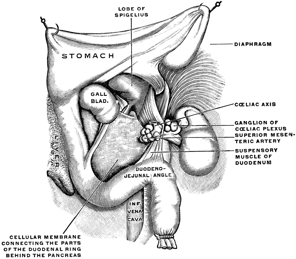
The ligament of Treitz is often said to consist of two parts [1, 20]: (1) a slip of striated muscle, derived from the diaphragm near the esophageal opening and ending in the connective tissue adjacent to the celiac arterial trunk, and (2) a fibromuscular band, containing smooth muscle or the so-called suspensory muscle of the duodenum, which originates from the duodenum and/or the.

Resultado de imagem para fascia de treitz Anatomy, Superior
The ligament of Treitz is commonly used as the point to differentiate the two. Bleeds proximal to the ligament are upper GI bleeds, and distal bleeds are lower GI bleeds. Categorization into one of the two groups is important as it directs the evaluation and management of the patient. [1] [2] [3]

Restoration bringing the AL to the ligament of Treitz. Download
Basics TerminologyIntroduction to the musculoskeletal systemIntroduction to the other systems Upper limb OverviewShoulder and armElbow and forearmWrist and handNerves and vessels Lower limb OverviewHip and thighKnee and legAnkle and footNerves and vessels Spine and back
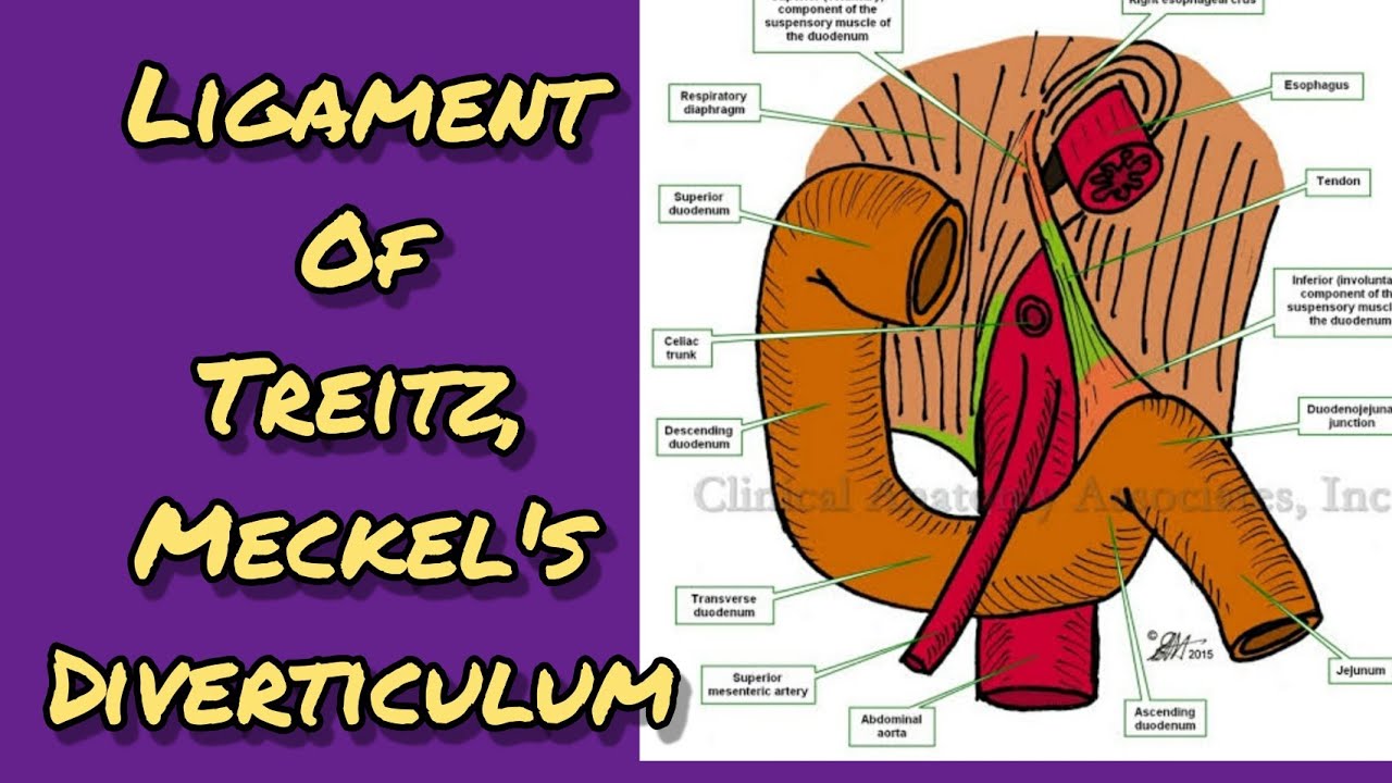
Ligament of Treitz , Meckel's diverticulum YouTube
Clinically the ligament of Treitz marks the border between the upper and lower gastrointestinal tract. Duodenum in a cadaver: It is easy to locate the duodenum and pancreas during a dissection. The duodenum follows a C-shaped trajectory around the head of the pancreas. Blood supply.

Ligamentum Treitz / Malrotation Springerlink / Treitz ligamenti
Abstract OBJECTIVE. The objective of this article is to discuss the anatomy, embryonic origin, normal variants, and various attachments of the ligament of Treitz. We also describe the pathologic.
Suspensory Muscle of the Duodenum ClipArt ETC
The ligament of Treitz is one of the frequently forgotten structures within the abdomen. It was named after the Austrian physician and anatomist Wenzel Treitz, who in 1853 first described the ligament as a thin, triangular, fibromuscular band extending from the upper surface of the duodenojejunal junction [1].

Represent the actual anatomy in our patient and the relationship of
The ligament of Treitz is a band of tissue in your abdomen (belly). It supports and anchors the small intestine and helps move its contents along. A birth defect involving the ligament can cause intestinal malrotation (twisting). Contents Overview Function Anatomy Conditions and Disorders Care Additional Common Questions Overview

suspensory muscle of the duodenum
The ligament of Treitz, also known as the suspensory ligament of the duodenum, is a double fold of peritoneum suspending the duodenojejunal flexure from the retroperitoneum. It is often used interchangeably with duodenojejunal flexure. Anatomy The ligament of Treitz comprises two parts: accessory muscle, also known as Hilfsmuskel 1
Ligament of Treitz Suspensory ligament of duodenum Kenhub
This flexure is attached to the posterior abdominal wall by a peritoneal fold called the suspensory muscle (ligament) of duodenum, also called the ligament of Treitz. The duodenum has four parts: superior (duodenal bulb/ampulla), descending, horizontal and ascending. Among several features of the duodenum, we'll list the two most important:

Ligament of Treitz GrepMed
The ligament of Treitz is an anatomical landmark used by anatomists and surgeons to denote the duodenojejunal junction and the point where the small intestine passes from retroperitoneal duodenum to intraperitoneal jeunum. Surgeons use the ligament of Treitz to measure the jejunum to decide where to perform an anastomosis.
Ligament of Treitz Suspensory ligament of duodenum Kenhub
The suspensory muscle of duodenum (also known as suspensory ligament of duodenum, Treitz's muscle or ligament of Treitz [1] ) is a thin muscle connecting the junction between the duodenum and jejunum (the small intestine 's first and second parts, respectively), as well as the duodenojejunal flexure to connective tissue surrounding the superior.

SUSPENSORY MUSCLE OF DUDENUM OR LIGAMENT OF TREITZ YouTube
The anatomic landmark that separates upper and lower bleeds is the ligament of Treitz, also known as the suspensory ligament of the duodenum. This peritoneal structure suspends the duodenojejunal flexure from the retroperitoneum.

Ligament of Treitz Diagram Quizlet
OBJECTIVE. The objective of this article is to discuss the anatomy, embryonic origin, normal variants, and various attachments of the ligament of Treitz. We also describe the pathologic processes that develop along the ligament of Treitz and the role of cross-sectional imaging in identifying these conditions. CONCLUSION.

Ligament Of Treitz Create your own flashcards or choose from millions
In medical terminology, the ligament of Treitz is referred to as duodenojejunal flexure. It is considered a tissue band in the abdomen. Different anatomists describe the ligament of Treitz as a suspensory muscle of the duodenum. Moreover, it arises from the connective tissue present near the stem level of celiac and superior mesenteric arteries.

Ligament of Treitz Anatomy, Relevance of Radiologic Findings, and
The ligament of Treitz is a thin muscular band that connects the duodenum and jejunum to the surrounding connective tissue. This serves as the marker for the transition from the duodenum to the jejunum. The jejunum is the second segment of the small bowel. It is approximately 100 cm long and is characterized anatomically by its circular.

Ligament of Treitz Anatomy, Relevance of Radiologic Findings, and
The ligament of Treitz is one of the frequently forgotten structures within the abdomen. It was named after the Austrian physician and anatomist Wenzel Treitz, who in 1853 first described the ligament as a thin, triangular, fibromuscular band extending from the upper surface of the duodenojejunal junction [ 1 ].